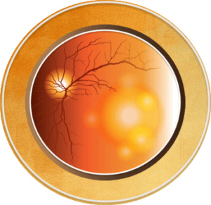
Stages of AMD
There are two types of age-related macular degeneration (AMD), usually described as either “dry” or “wet” (see Figure below).1,2 In the dry type of AMD, areas of the macula get thinner and deposits of fats and protein, called drusen, develop under the tissues of the retina.1,3 In the wet type of AMD, abnormal blood vessels grow under the tissues of the retina.1,2 These abnormal blood vessels can leak blood or other fluid, causing scarring of the macula.1 Both types of AMD can cause severe vision loss, affecting central vision but sparing the peripheral (side) vision.1,4
- “Dry” (atrophic) AMD: Approximately 80% of people with AMD have the dry form, which can slowly progress to late- (geographic) or end-stage disease2
- “Wet” (neovascular) AMD: Although the wet form is less common, vision loss can be faster and more severe; about 90% of cases of legal blindness caused by AMD are due to this form2,5

Although AMD has 2 types, it also can be separated into 3 stages2, 7-12:
- Early AMD: Usually characterized by small to medium-size drusen
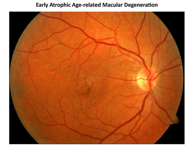
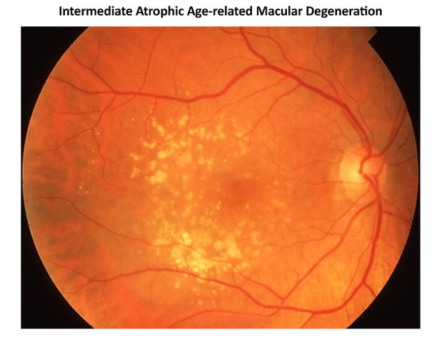
- Intermediate AMD: More, larger drusen accumulate, and some areas of retinal tissue thinning develop around the macula
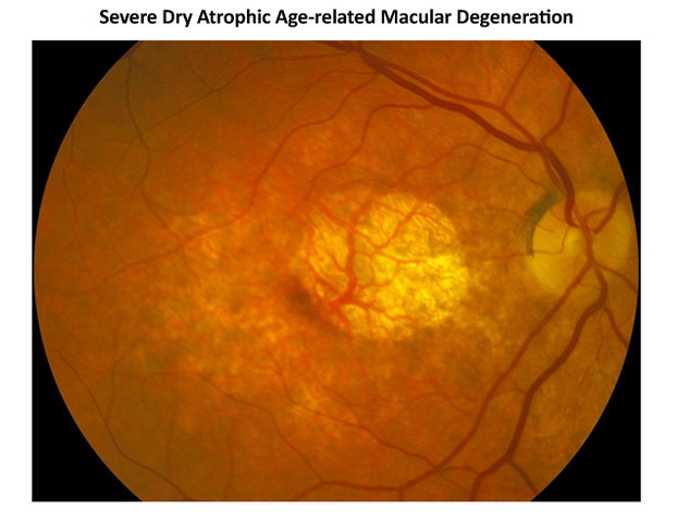
- Severe AMD can be characterized in 2 ways:
- Severe dry AMD (also called geographic atrophy), with retinal tissue thinning involving the center of the macula
- Wet AMD (also called neovascular AMD)
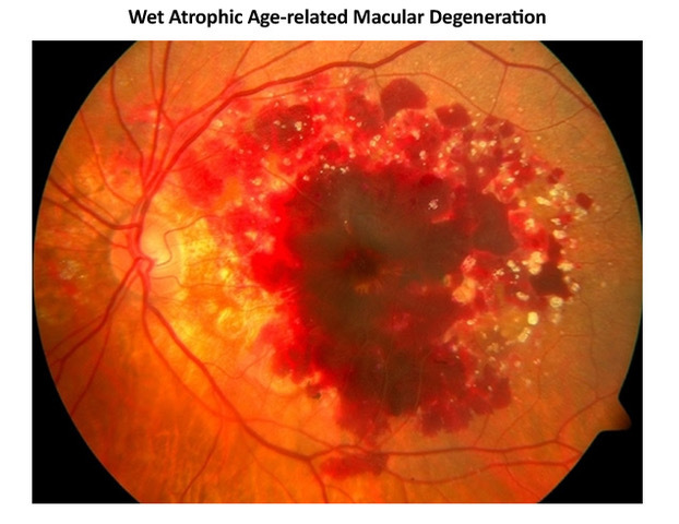
Although people with early and intermediate stages of AMD rarely have symptoms, people with severe or wet AMD usually do experience symptoms.13 Earlier diagnosis and appropriate treatment helps maintain better vision.14
References
- American Academy of Ophthalmology (AAO). What is macular degeneration? 2021. https://www.aao.org/eye-health/diseases/amd-macular-degeneration.
- AAO. Age-related macular degeneration Preferred Practice Pattern®. Ophthalmology. 2020;127:P1-P65. https://www.aao.org/preferred-practice-pattern/age-related-macular-degeneration-ppp.
- AAO. What are drusen? 2021. https://www.aao.org/eye-health/diseases/what-are-drusen.
- Klein R, Klein BE, Linton KL. Prevalence of age-related maculopathy; the Beaver Dam Eye Study. Ophthalmology. 1992;99:933-943.
- Shaw PX, Stiles T, Douglas C, et al. Oxidative stress, innate immunity, and age-related macular degeneration. AIMS Mol Sci. 2016;3:196-221.
- Shutterstock.com. Age-related macular degeneration. Item 83736805. https://www.shutterstock.com/image-illustration/eye-condition-macular-degeneration-83736805.
- Ferris FL 3rd, Wilkinson CP, Bird A, et al. Clinical classification of age-related macular degeneration. Ophthalmology. 2013;120:844-851.
- American Society of Retina Specialists (ASRS). The Foundation Retina Health Series. Age-related macular degeneration. 2022. https://www.asrs.org/patients/retinal-diseases/2/age-related-macular-degeneration.
- Diseases at the back of the eye. Comm Eye Health J. 2014;27:70-71.
- Cook GR. Retina Image Bank. 2019;29839. https://imagebank.asrs.org/file/29839/atrophic-amd-with-soft-drusen.
- Huang SS. Age-related macular degeneration – geographic atrophy. Retina Image Bank. 2013;6298. https://imagebank.asrs.org/file/6298/age-related-macular-degeneration-geographic-atrophy.
- Kelly MP. Retina Image Bank. 2012;729. https://imagebank.asrs.org/file/729/age-related-macular-degeneration.
- Ayoub T, Patel N. Age-related macular degeneration. J R Soc Med. 2009;102:56-61.
- Rauch R, Weingessel B, Maca SM, Vecsei-Marlovits PV. Time to first treatment: the significance of early treatment of exudative age-related macular degeneration. Retina. 2012;32:1260-1264.
All URLs accessed 2/8/22.
